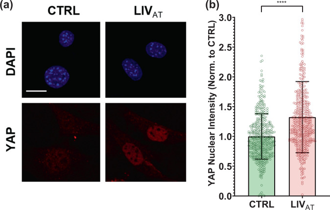
Fig. 1. Acute LIVAT application increases nuclear YAP levels.
a MSCs were subjected to LIVAT and stained with DAPI (blue) and YAP (red). Confocal images indicated increased nuclear YAP levels following acute LIVAT applied as five 20 min vibration periods separated by 1 h. b Quantitative analysis of confocal images showed a 32% increase of nuclear YAP in LIVAT samples compared to controls. n > 400/grp, group comparison was made a Mann–Whitney U-test, ****p < 0.0001. Error bars represent standard deviation. Scale bar: 10 μm. Full statistical details were provided in Supplementary Table 4.

