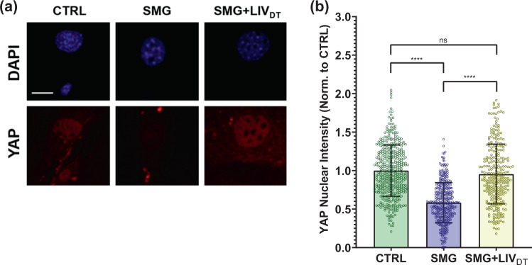
Fig. 2. Basal nuclear YAP levels decreased by SMG were rescued by LIVDT.
a MSCs were subjected to SMG, and SMG + LIVDT over 72 h period and stained with DAPI (blue) and YAP (red). b Quantitative analysis showed a 42% decrease in nuclear YAP levels in the SMG group compared to control levels. The SMG + LIVDT group showed a 67% increase in nuclear YAP when compared to the SMG group. There was no statistically significant difference between CTRL and SMG + LIVDT groups. n > 100/grp. Group comparisons were made via Kruskal–Wallis test followed by Tukey multiple comparison, ****p < 0.0001. Error bars represent standard deviation. Scale bar: 10 μm. Full statistical details were provided in Supplementary Table 5.

