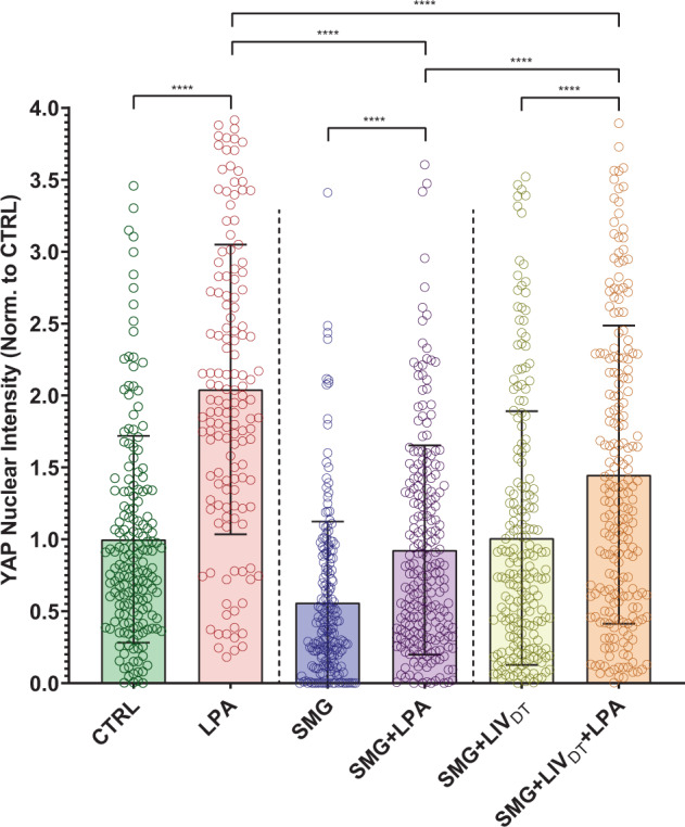
Fig. 6. LPA-induced YAP nuclear entry decreased by SMG was alleviated by daily LIVDT application.

MSCs were subjected to SMG, and parallel SMG + LIVDT over 72 h period at the end of 72 h, samples were treated with either LPA (50 µM) or DMSO. Quantitative analysis of confocal images revealed that LPA addition increased nuclear YAP levels by 105%, 67%, and 43% in the CTRL, SMG, and SMG + LIVDT when compared to DMSO controls. When compared to nuclear YAP intensity of the LPA treatment alone, SMG + LPA and SMG + LIVDT + LPA samples were 55% and 29% lower, respectively. YAP nuclear levels in SMG + LIVDT + LPA remained 70% higher than SMG + LPA group. n > 100/grp. Group comparisons were made via Kruskal–Wallis test followed by Tukey multiple comparison, ****p < 0.0001. Error bars represent standard deviation. Scale bar: 10 μm. Full statistical details were provided in Supplementary Tables 8.
