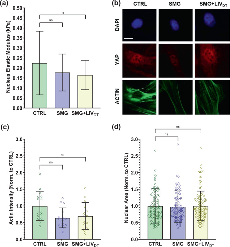
Fig. 7. MSC stiffness and structure remain intact under SMG and SMG + LIVDT treatments.
MSCs were subjected to SMG and parallel SMG + LIVDT over a 72 h period. a Compared to CTRL samples, AFM measurement of the elastic moduli of SMG and SMG + LIVDT-treated MSCs revealed apparent decreases in elastic modules that were 21 and 27% below control levels, measured differences were not statistically significant. n = 10/grp. b Quantification of confocal images shows that c mean F-actin intensity of SMG and SMG + LIVDT-treated MSCs revealed a decrease of 36 and 30% below control levels, measured differences were not statistically significant. n = 15/grp. d No significant effects of either SMG or LIVDT treatment on the average nucleus size were found. n > 100/grp. Group comparisons were made via Kruskal–Wallis test followed by Tukey multiple comparison. Error bars represent standard deviation. Scale bar: 10 μm. Full statistical details were provided in Supplementary Tables 9–11.

