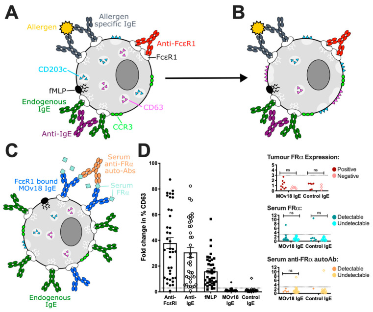
Figure 5.
Basophil activation test (BAT). (A) Basophils can be identified in unfractionated whole blood samples by cell surface markers, such as CCR3 and CD203c. (B) Surface expression of CD63 and CD203c is upregulated in activated basophils through the fusion of intracellular vesicles, via both IgE- (anti-FcεRI, anti-IgE and allergen cross-linking of specifc IgE) and non-IgE-mediated (fMLP) stimuli. Adapted from Bax et al. [11]. (C) Circulating FRα anti-FRα autoantibodies found in ovarian cancer patient sera may form immune complexes with MOv18 IgE triggering basophil activation. (D) Ex vivo basophil activation (>3.0 fold change in % CD63 expression) was measured following incubation with anti-FcεR1, anti-IgE, fMLP stimulation, but not triggered by MOv18 and control IgE antibodies, in all but one patient. Activation by MOv18 IgE, or lack thereof, was irrespective of patient tumour FRα expression, or the presence of FRα and anti-FRα autoantibodies in patient sera. Adapted from Bax et al. [79].

