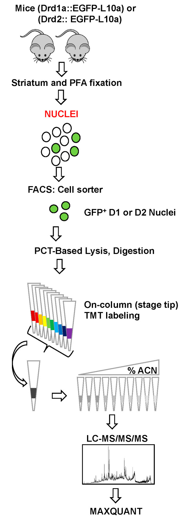
Figure 1.

Proteomics workflow: Nuclei from striatal tissue from Drd1a:EGFP-L10a or Drd2:EGFP-L10a transgenic mice were sorted into microtubes. Lysis and trypsin digestion were then carried out using a PCT-MicroPestle (Pressure-cycling technology). After desalting and on-column TMT labeling in C18 StageTips, the samples were mixed and then subjected to pH 10 fractionation on sulfonated divinylbenzene (bSDB) packed StageTips. The resulting fractions were then analyzed by LC-MS/MS/MS with the raw data being processed by MaxQuant.
