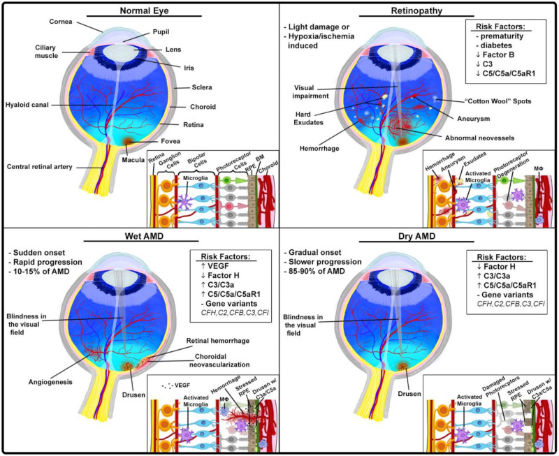
Figure 3.
Ocular pathologies with complement involvement. Anatomy and histology of normal eye (upper left); proliferative retinopathies (upper right): the process of neovascularization leads to formation of blood vessels that are fragile and leaky, increased permeability or rupture of newly formed blood vessels and subsequent hemorrhage cause impairment or loss of vision, in mouse models of these retinopathies, complement seems to have a protective role; wet AMD (lower left) and dry AMD (lower right): in both forms, excessive complement activation and subsequent generation of complement effectors (C3a and C5a) has been implicated; frequently AMD is linked to FH polymorphism; in wet AMD, VEGF-driven neovascularization, possibly also regulated by complement, leads to severe impairment or loss of vision.

