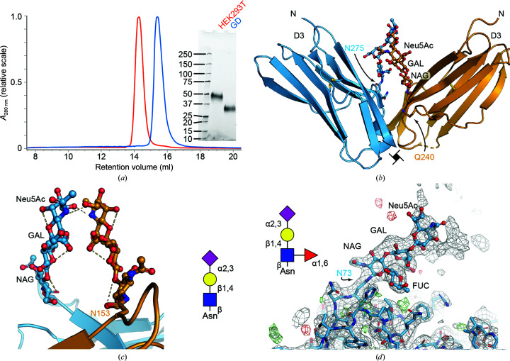
Figure 2.
Structural analysis of CSF-1RD1–D3 derived from HEK293 GD. (a) Comparison of HEK293T and HEK293 GD-derived CSF-1RD1–D3; the inset shows reduced SDS–PAGE analysis of the purified proteins. (b) Van der Waals contacts between GD-type glycans on Asn275. (c) Crystal-packing contacts between symmetry-related glycans. The inset shows a schematic representation of the glycan. (d) The GD-type glycan on Asn73 is α-1,6-fucosylated. The 2mF o − DF c electron-density map is shown as a gray mesh. Residual positive and negative mF o − DF c electron-density maps (contoured at ±3σ) are shown in green and red, respectively.

