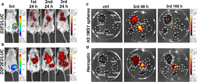
Fig. 2.
Representative images indicating the biodistribution of functionalized silk spheres in Her2(+) and Her2(−) mouse orthotopic breast cancer models. Biodistribution of H2.1MS1 spheres over time after intravenous injection of BALB/c mice, which developed a Her2(−) D2F2 tumors and b Her2(+) D2F2E2 tumors. Biodistribution of c H2.1MS1 spheres and d Herceptin 48 and 168 h after the 3rd intravenous injection in mice that developed D2F2E2 tumors. Organs and tumors were excised and imaged. Signal detection was performed by using the IVIS Spectrum system to estimate the luminescence intensity of the tumors and the fluorescence intensity of the ATTO647N-labeled H2.1MS1 spheres and Herceptin at wavelengths of 640/680 nm

