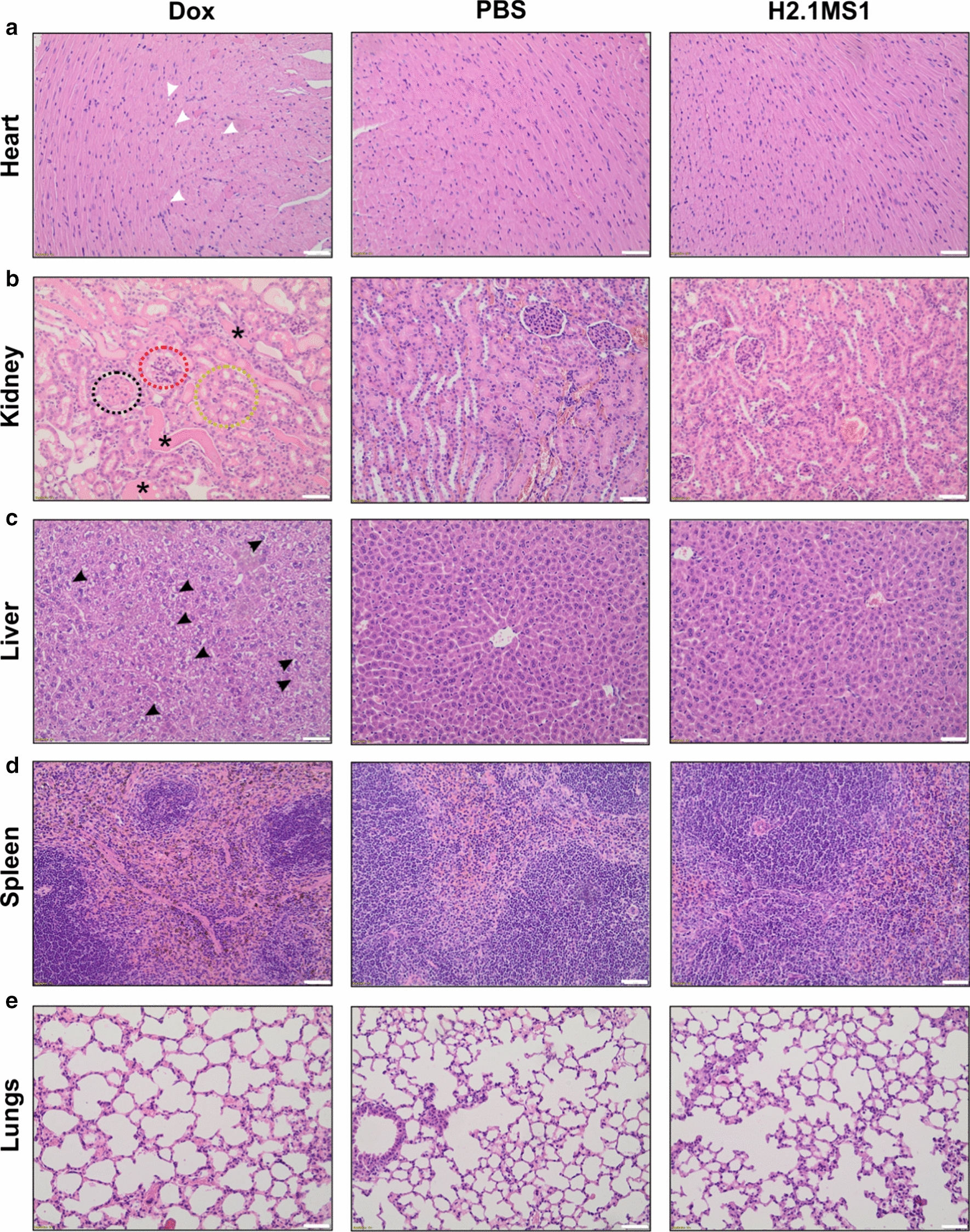
Fig. 5.

H&E staining of FFPE sections of organs collected after treatment. Her2(+) D2F2E2 tumor-bearing mice were injected intravenously with free Dox, PBS and Dox-loaded H2.1MS1 spheres according to the schedule presented in Fig. 4a. Organs such as the heart, kidney, liver, spleen, and lungs were excised on the 20th day, and the samples were stained with H&E. White arrows indicate small vacuoles in cardiomyocytes, and black arrows indicate vacuolar degeneration in the liver. The circles point out glomerular hyalinization (black), vacuolization of glomeruli (red), and hyaline droplets degeneration (yellow), and the asterisks indicate renal tubular dilatation with protein casts. Scale bar: 100 μm
