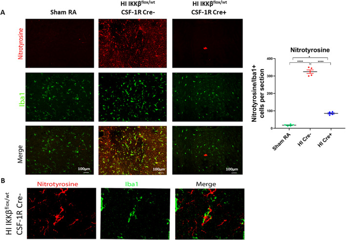
Fig. 7.
NF-κB activation increases nitrative stress, causing injury to the vulnerable oligodendroglia. a Immunohistochemistry of coronal sections of the periventricular brain area of postnatal day 15 pups. Top panel shows Nitrotyrosine (marker of nitrative stress) in red. Middle panel shows Iba1 (microglial marker) in green. Lower panel shows Co-localization in yellow nitrotyrosine secreted by microglia. Quantification of nitrotyrosine/Iba1 per section. Scale bar = 200 μm. N = 5 animals/group and 4 sections/animal. Scatter dot plot showing mean ± SE. *P < 0.05, ****P < 0.0001. b Higher magnification in the HI IKKβflox/wt CSF-1R Cre− (HI Cre−) group showing the co-localization of nitrotyrosine to Iba1 indicating that microglia is source of the nitrative stress (nitortyrosine)

