Fig. 5.

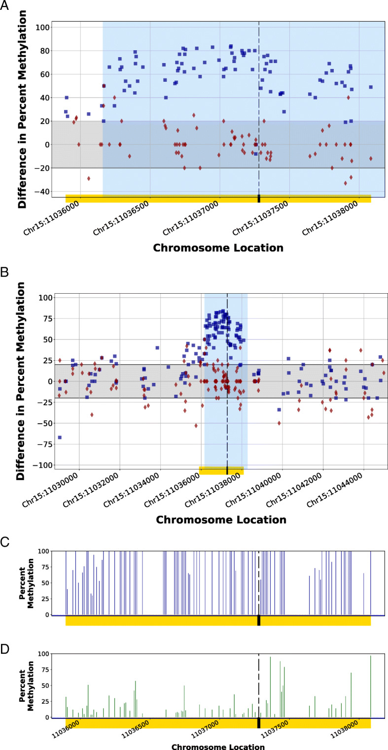
Methylomic difference of a completely edited CpG island within the genome of the HDR1 Animal using homology-directed repair in a CRISPR-mediated edit. Homology arms of a synthetic DNA fragment (region shaded in blue) in a homology-directed repair of a CRISPR-mediated genomic cut spanned a CpG island (CGI; Chr15:11,035,894–11,038,084; yellow bar) within the genome of HDR1 Animal, resulting in a destabilized methylation pattern (blue squares) as compared to the Control 1 Animal methylation patterns at the same location (red diamonds). Figure 4a illustrates the localized methylation variance introduced at the CGI, while Fig. 4b illustrates the comparison of the modified region to flanking endogenous genomic regions by displaying the variance of methylation patterns for 7000 bp upstream and downstream of the CGI. The methylation patterns of regions not influenced by the incorporation of donor DNA during the CRISPR edit displayed a methylation pattern similar to the control. The gray band indicates considered biological epigenetic variance (+/− 20% change). The dashed line indicates the protospacer adjacent motif (PAM) location of the targeted CRISPR cut site. Synthetic homology arms corresponded to genomic sequence 1000 bp upstream and downstream of the PAM site. The comparison of the percent methylation observed at the localized CGI region for the CRISPR-edited animal (Fig. 4c) and the unedited control animal (Fig. 4d) demonstrate the introduced methylation variance as a result of the introduction of the donor DNA. Blue squares (■) indicate the percent differences in CpG methylation for HDR1 Animal from Control 2 Animal at given chromosome locations. Red diamonds (♦) indicate the percent differences in CpG methylation for Control 1 Animal from Control 2 Animal at given chromosome locations
