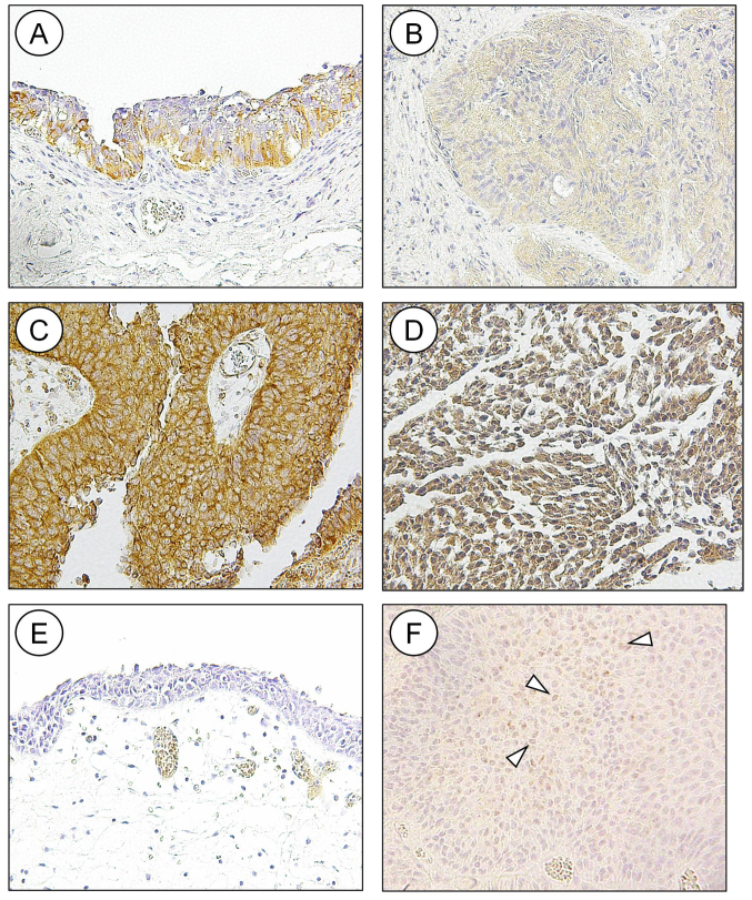
Figure 1.
Immunohistochemical analysis of HDAC6 and PD-L1 in normal urothelium and UC. (A) HDAC 6 was detected in the cytoplasm of normal urothelium. Staining for HDAC6 was either (B) low or (C and D) high and was detected in the (C) cytoplasm or (D) nucleus and cytoplasm of UC. Membranous staining of PD-L1 was not detected in (E) normal urothelium; however, it was detected in (F) UC cells (indicated by the arrowheads). UC, urothelial cancer; PD-L1, programmed death-ligand 1; HDAC, histone deacetylase.

