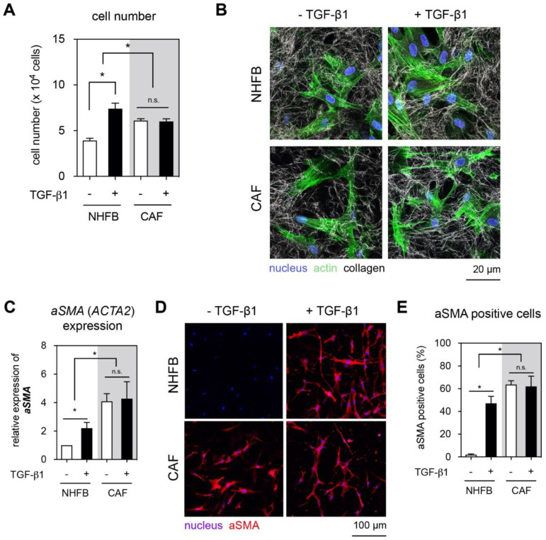
Figure 1.
Effect of TGF-β1 on proliferation, aSMA expression, and matrix remodeling of NHFB and CAF. NHFB and CAF were cultured on 3D collagen matrices in the presence and absence of TGF-β1 for 7 days. Both cells were then characterized in terms of proliferation, aSMA expression, and matrix remodeling. (A) Quantitative analysis of cell number using commercial WST-1 assay. (B) Representative confocal images of NHFB and CAF. Cells were stained with DAPI and Phalloidin conjugated with Alexa Fluor 488 to visualize nucleus and actin, respectively. Collagen fibrils were imaged in the reflection mode. (C) The expression of aSMA was analyzed using qPCR and was normalized to NHFB cultured in the absence of TGF-β1. (D) Immunocytostaining of aSMA and (E) quantitative analysis of number of aSMA positive cells by manual count. Data are represented as mean ± SD; *—significance level of p < 0.05; n.s.—not significant. All quantitative experiments were performed at least in 3 replicates with fibroblasts and CAF from 3 different donors for each condition.

