Figure 5.
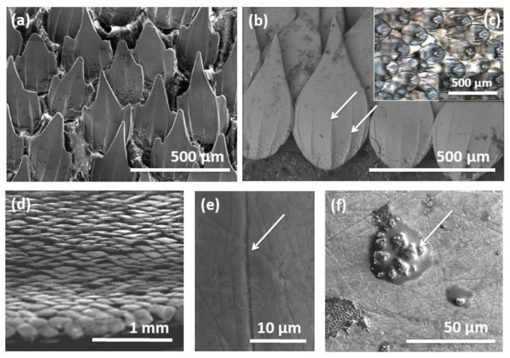
The shape and structure of dermal denticles from the small catshark, Scyliorhinus canicula (examined using electron microscopy). ((a,b) scale bar = 500 µm) Variations in the shape of dermal denticles observed from skin samples in different body locations in one individual specimen of this species. Similar structures to the microscopic ridges on the surface of individual denticles (as indicated by arrows in (b), scale bar = 500 µm) are thought to be responsible for drag reduction in sharks. It is interesting to observe the degree of overlap between denticles (inset (c), scale bar = 500 µm) and how the denticles extend and completely envelope the shark, including the trailing edges of fin surfaces (d), scale bar = 1 m)m. The role these structures play in both drag reduction and in antifouling is still of keen research interest. An interesting observation from close examination of denticle surfaces in S. canicula is that the upper surface of individual denticles are often scarred and grooved with microscopic ridges (e), scale bar = 10 µm) perhaps from contact with other sharks or other behaviours that remove attached organisms, although fouling may be observed in some regions (f) scale bar = 50 µm.

