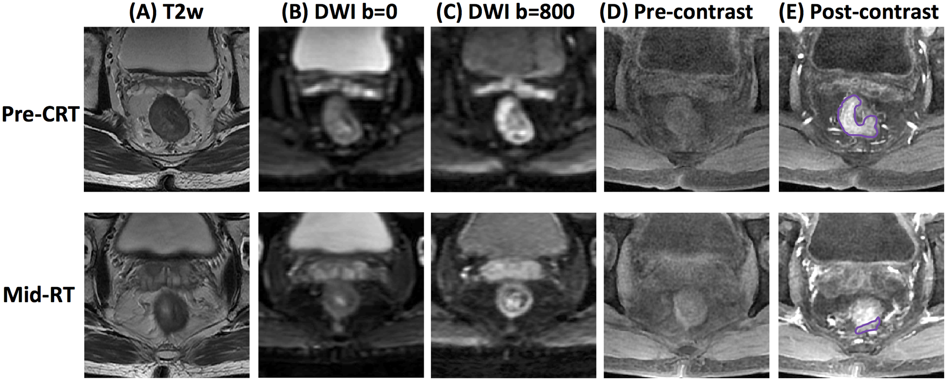
Figure 1.

MR images of a 51-year-old male with low-rectum cancer at stage of cT3N+M0 taken pre-treatment (top row) and mid-RT (bottom row). (A) T2-weighted image, (B) the diffusion-weighted image with b=0 s/mm2, (C) the diffusion-weighted image with b=800 s/mm2, (D) L1 pre-contrast image, (E) L2 post-contrast image taken at 15 seconds after injection. This patient achieved pCR after completing the entire course of CRT.
