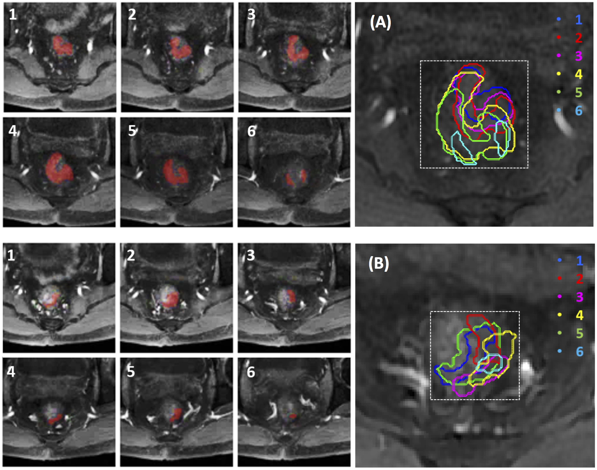
Figure 2.

Determination of smallest bounding box on pre-treatment MRI (A, top panel) and mid-RT MRI (B, bottom panel) of a 56-year-old male with mid-rectum cancer at stage of cT3N+M0. Tumor ROI (red) outlined on tumor-containing MR slices (1–6) are stacked on a projection view to determine the smallest square bounding box.
