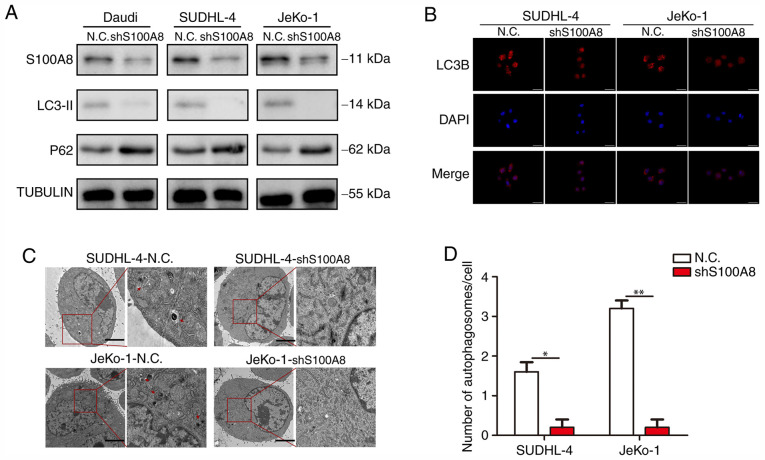
Figure 4.
S100A8 increases autophagy in BCL cells. (A) Western blotting for LC3-II and P62 in BCL cells was performed following S100A8 interference. (B) Representative images of LC3-positive puncta in BCL cells following S100A8 interference. (C) Transmission electron microscopy of BCL cells following S100A8 interference. Autolysosomes are indicated by arrowheads. Scale bars, 5 µm. (D) Quantification of detectable autolysosomes in BCL cells following S100A8 interference. **P<0.01 and *P<0.05. BCL, B-cell lymphoma; LC3, microtubule-associated protein 1A/1B-light chain 3.

