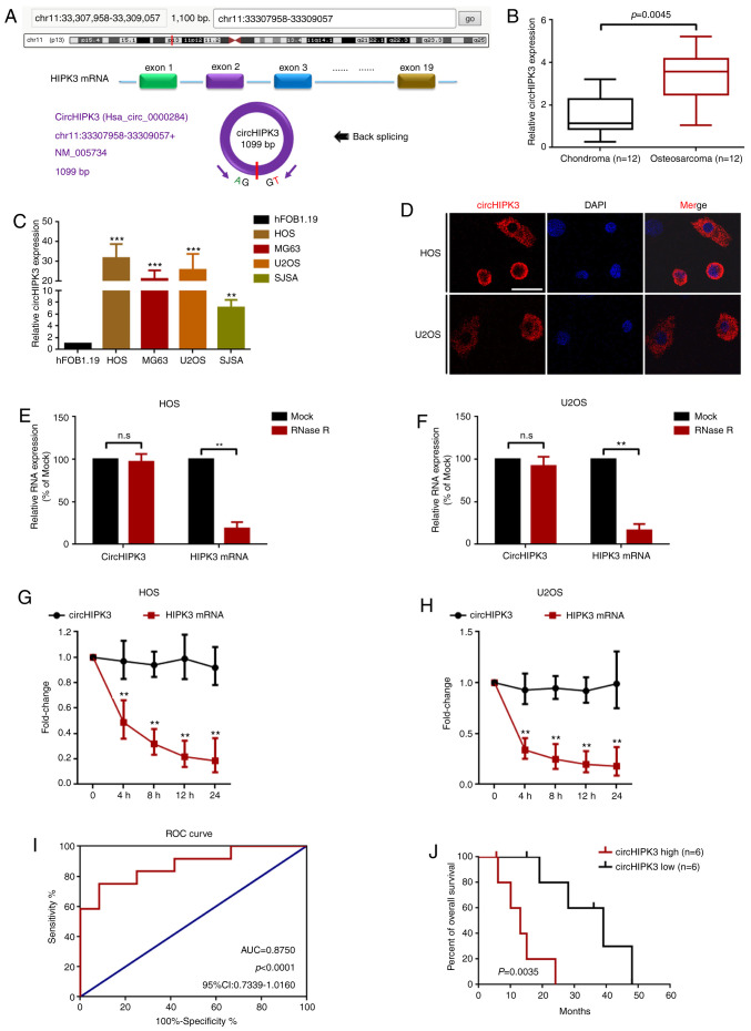
Figure 1.
circHIPK3 is relatively highly expressed in OS cells, and predominantly localized in the cytoplasm. (A) circHIPK3 was derived from exon 2 of linear HIPK3 mRNA as illustrated. (B) circHIPK3 expression in 12 pairs of OS and chondroma tissue samples was determined using RT-qPCR. (C) circHIPK3 expression in four OS cell lines and in the normal osteoblast hFOB1.19 cell line was measured using RT-qPCR. **P<0.01 and ***P<0.001 vs. hFOB1.19. (D) circHIPK3 was mainly located in the cytoplasm according to a fluorescence in situ hybridization assay. Magnification, ×400; scale bar, 20 µm. An RNase R assay was performed to determine the stability of circHIPK3 in (E) HOS and (F) U2OS cells. **P<0.01. An actinomycin D assay was performed to check the stability of circHIPK3 in (G) HOS and (H) U2OS cells. **P<0.01. (I) Clinical values of circHIPK3 in OS were analyzed using an ROC curve (AUC, 0.8750; P<0.0001). (J) High expression levels of circHIPK3 were associated with shorter overall survival of patients with OS. n=6 for each group. P=0.0035. All data are presented as the mean ± SD of three independent experiments. AUC, area under the curve; circ, circular RNA; HIPK3, homeodomain interacting protein kinase 3; n.s, not significant (P>0.05); OS, osteosarcoma; ROC, receiver operating characteristic; RT-qPCR, reverse transcription-quantitative PCR.

