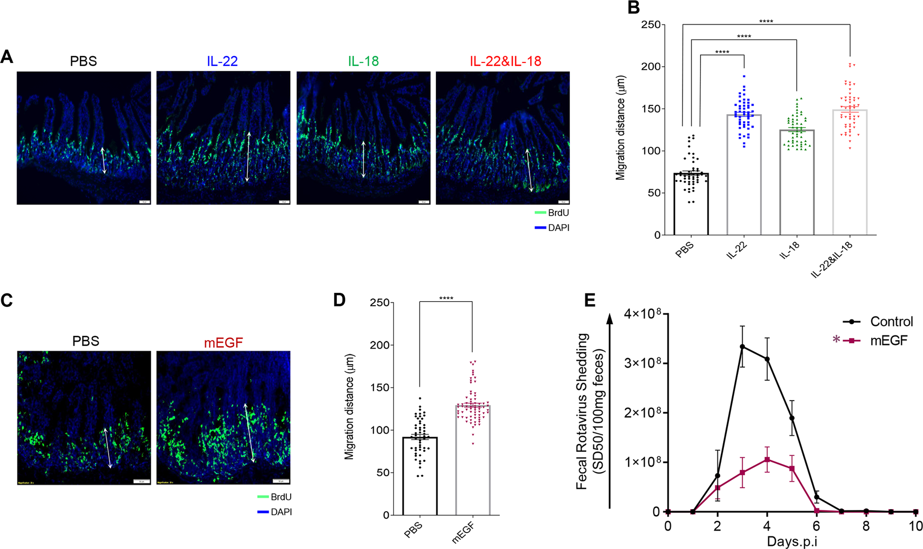
Fig. 3. Accelerated proliferation rate and migration levels of IEC are correlated with debilitation of mRV infectivity.

Mice were i.p injected with PBS, IL-22, (10 μg) IL-18 (2 μg), both cytokines, or murine epidermal growth factor (mEGF). 1h later, mice were administered BrdU. Mice were euthanized 16-hour post-BrdU administration, and BrDU was visualized (A and C) and migration was measured (B and D) by microscopy and image analysis, respectively. Images shown in A and C are representative. For B and D, sections were scored at least from 50 villus per group of mice (n = 5). Distance of the foremost migrating cells along the crypt-villus axis were measured with ImageJ software. Results are presented as mean ± SEM. Statistical significance was evaluated by Student’s t-test (**** denotes P < 0.0001). (E) Mice were i.p injected with PBS or EGF (10 μg) murine EGF two hours prior to, or 2, 4, 6, or 8 days after oral inoculation with mRV. Fecal RV levels were measured over time by ELISA. Data are mean ± SEM, n=5 * indicates significantly different from PBS by two-way ANOVA, P < 0.0001.
