
Figure 1. RACK1 Interacts with NLRP3 and NEK7.
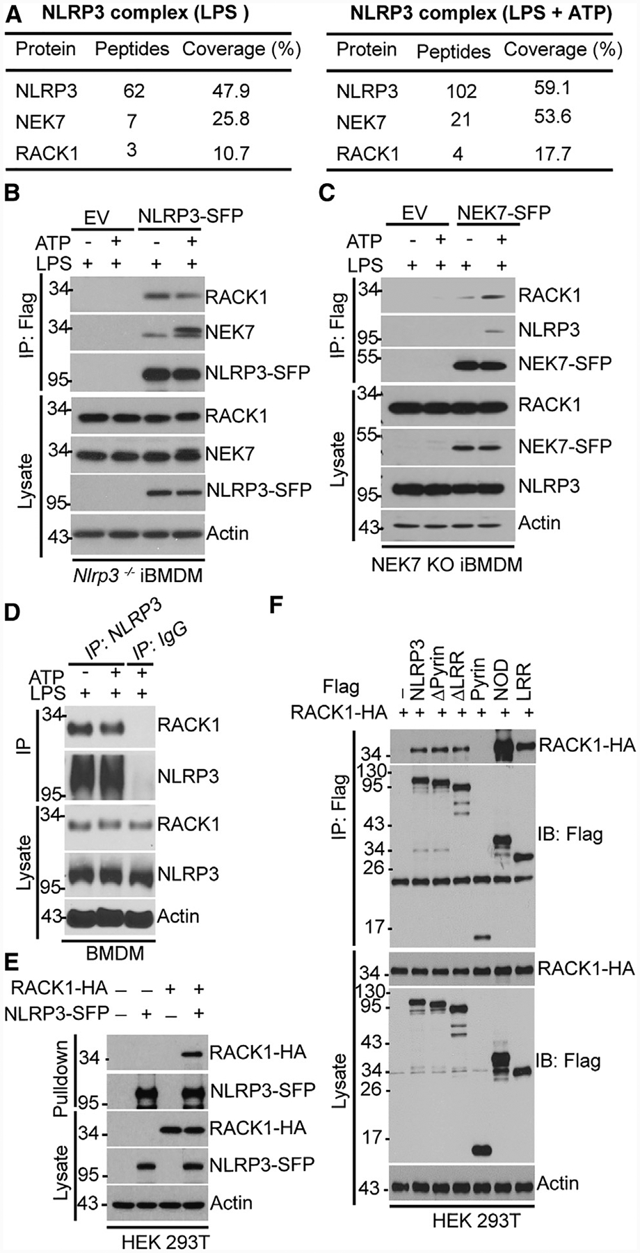
(A) Mass spectrometry analysis of NLRP3, RACK1, and NEK7 peptides after purification of NLRP3 complexes.
(B and C) Tagged NLRP3 (NLRP3-SFP) or NEK7 (NEK7-SFP) was immunoprecipitated (IP) with anti-FLAG antibody from iBMDM treated with LPS (200 ng Ml−1, 4 h) alone or with LPS plus ATP (5 mM, 30 min) and was immunoblotted with the indicated antibodies. EV, empty vector.
(D) BMDM was stimulated with LPS (200 ng mL−1, 4 h) alone or with LPS plus ATP (5 mM, 30 min). Cell lysates were immunoprecipitated and immunoblotted with the indicated antibodies.
(E) NLRP3-SFP was co-expressed with HA-tagged RACK1 in HEK293T cells, pulled down, and analyzed by immunoblotting. HA, hemagglutinin.
(F) FLAG-tagged, full-length or truncated NLRP3 was co-expressed with HA-tagged RACK1 in HEK293T cells, immunoprecipitated, and analyzed by immunoblotting. (Δpyrin, pyrin domain deleted; ΔLRR, leucine-rich repeats deleted; pyrin, pyrin domain only; NOD, NOD domain only; LRR, leucine-rich repeat only). Results are representative of three independent experiments.