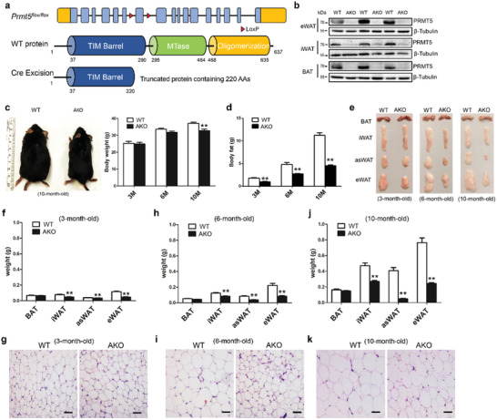
Figure 1.

Adipocyte‐specific knockout of Prmt5 (Prmt5AKO) leads to loss of fat mass in white adipose tissues. a) Targeting strategy for conditional knockout of Prmt5. Upper: Prmt5 gene structure showing exons (blue boxes), LoxP insertions (red triangles) and 5′ and 3′ untranslated regions (orange boxes). Middle: PRMT5 protein domain structure with amino acids numbers labeled. MTase is the catalytic domain. Lower: Cre‐mediated excision of LoxP‐flanked exon results in reading frame shift and a premature translational stop codon, generating a truncated protein containing only partial of the TM barrel domain. b) Efficient reduction of PRMT5 protein levels in epididymal White Adipose Tissue (eWAT) and inguinal White Adipose Tissue (iWAT), and brown adipose tissue (BAT) of the 2 month old male Prmt5AKO (AKO) mice. c) Representative images of male WT (Prmt5flox/flox) and Prmt5AKO mice at 10 month old (left panel), showing progressive reduction in body weight of the Prmt5AKO mice relative to WT mice (right panel). d) Prmt5AKO leads to progressive loss of total body fat, relative to fat mass in WT mice. c,d) n = 8, 6, and 4 pairs of male mice for 3‐, 6‐, and 10‐month‐old, respectively. e) Representative images of BAT and WAT depots from male mice showing progressive mass reduction of Prmt5 KO WAT with age. f,h,j) Weights of various BAT and WAT (iWAT, eWAT and anterior subcutaneous White Adipose Tissue, asWAT) depots from male mice at different ages, as shown in (e). g,i,k) H&E staining of eWAT sections from WT (Prmt5flox/flox) and Prmt5AKO male mice at 3, 6, and 10month old. Scale bar: 50 µm. Data represent mean ± s.e.m. (t‐test: **p < 0.01).
