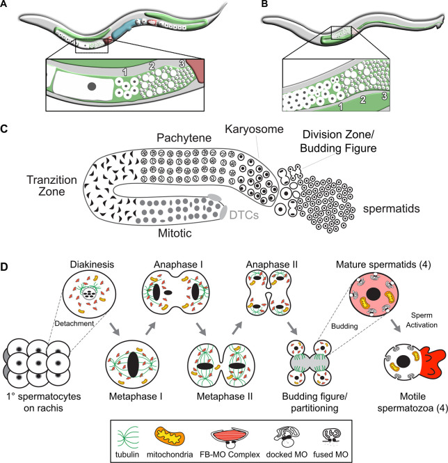
Fig. 1.
Overview of C. elegans spermatogenesis. (A,B) Cartoons depicting a young adult C. elegans hermaphrodite (A) and male (B), and their respective germlines. The hermaphrodite germline (A) is transitioning from spermatogenesis to oogenesis. The enlarged views highlight the linear arrangement of the primary spermatocytes (1), residual bodies (RBs) (2) and mature haploid spermatids (3). (C) Stylized cartoon of a surface view of the male germline highlighting its overall linear organization. Mitotic proliferation of the germline stem cells is maintained by two somatic distal tip cells (DTCs) that form the germ cell niche. The early events of meiotic prophase, including homolog pairing and formation of the synaptonemal complex, occur in the transition zone. Following an extended pachytene stage, spermatocytes enter a karyosome stage before mature spermatocytes detach from the syncytial germline and divide meiotically. The first meiotic division is often incomplete, leaving secondary spermatocytes linked by a cytoplasmic connection. Following anaphase II, the spermatocytes morph into budding figures that split into residual bodies and haploid spermatids. (D) Details of the meiotic divisions and post-meiotic partitioning event. Once spermatocytes detach from the germline syncytium, they pass through a brief diakinesis stage before undergoing nuclear envelope breakdown and initiating meiotic divisions. During the post-meiotic partitioning event, microtubules become acentrosomal and localize to the developing residual body (Winter et al., 2017). Components retained in the spermatids include fibrous body-membranous organelles (FB-MO), mitochondria, chromatin and centrioles. Components discarded within the RB include the tubulin, actin, endoplasmic reticulum and ribosomes of the cell; mature sperm are thus both transcriptionally and translationally inactive. Following separation from the RB, FBs disassemble and release unpolymerized MSP and the MOs dock with the plasma membrane. Males store sperm in this inactive spermatid state. During spermatid activation, MOs fuse with the plasma membrane and unpolymerized MSP localizes to the pseudopod, where it forms fibers that are required for spermatozoon motility.

