Fig. 3.

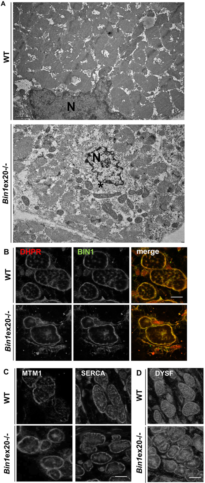
Alterations in intracellular organization and triads upon BIN1 defect. (A) Electron microscope visualized ultrastructure showed a general disorganization of muscle from newborn Bin1ex20−/− mice with a central collapse of nuclei (N), surrounded by an area devoid of myofibrils and filled with mitochondria and amorphous materials (*). (B,C) BIN1 detected with a pan-isoform antibody colocalized with the T-tubule marker DHPR in newborn muscle fiber (transversal view). Markers of T-tubules (DHPR in B), junctional sarcoplasmic reticula (MTM1 in C) longitudinal sarcoplasmic reticula (SERCA in C) were collapsed to the center of myofibers in the Bin1ex20−/− mice. (D) The marker of premature T-tubules (dysferlin) was collapsed to the center in Bin1ex20−/− mice. Of note, muscle fibers in wild-type newborns are usually less polygonal than in adults. Immunolabeling of 8 µm transversal section. Scale bars: 1 µm (A); 10 µm (B-D).
