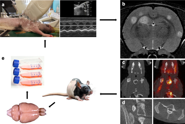
Fig 1. Establishment of the MDA-MB-231br/eGFP subpopulations.
a. Intracardiac injection of the MDA-MB-231br/eGFP cancer cells using ultrasound guidance. b-d. Follow-up of metastasis development with multimodal imaging. Preclinical 7 T MRI for assessment of brain metastases (b), [18F]FDG PET/CT for visualization of extracranial metastases (c), and high-resolution CT for the detection of bone metastases (d). e. Isolation of the MDA-MB-231br/eGFP cancer cells from the brain metastatic lesions followed by cell culture. This procedure was repeated five times.

