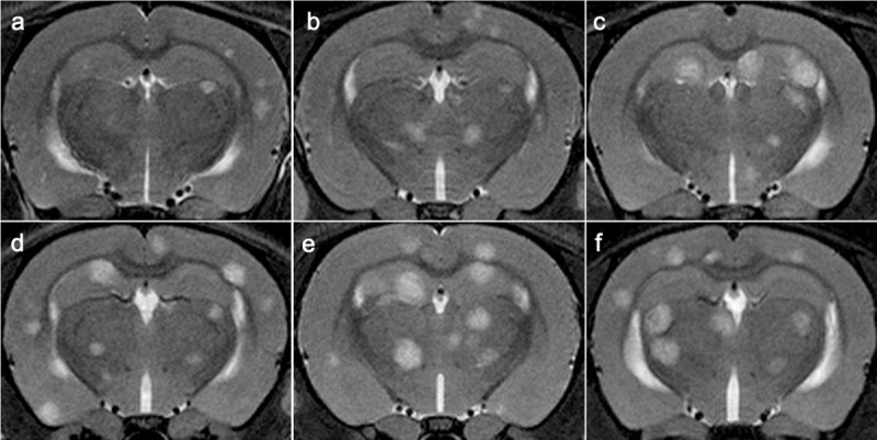
Fig 2. MR imaging of brain metastasis development per passage.

a–f. In vivo serial T2w MRI scans showing an increase of brain metastasis development per passage. T2w MR images from a representative case for P1 (a), P2 (b), P3 (c), P4 (d), P5 (e), P6 (f).
