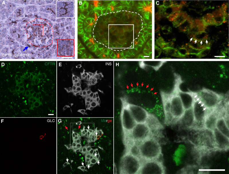
Fig 1. CFTR is expressed in human islets.
A-C. Immunohistochemistry (A) and immunofluorescence (B-C) images of human pancreas (normal tissue from an 11-month-old control) immunostained by using CFF-217 antibody (Table 1). The islet shown in A, encircled by a dashed red trace, contains endocrine cells stained by the antibody, which is shown in the magnified red square in the right bottom corner of the figure. CFTR-positive pancreatic duct cells are shown within the white squares. Shown in B is CFTR immunoreactivity in human exocrine and endocrine cells. The edges of all cells were immunolabeled by using E-cadherin antibodies (Table 1). The islet is encircled by a dashed trace and the square represents the area magnified and shown in C. D-H. In situ hybridization of normal human pancreas tissue by using fluorophore-labeled RNA probes directed against CFTR (green, D), insulin (INS, white, E) and glucagon (GLC, red, F) transcripts. Red and white arrows in H, a magnification of G, indicate specific CFTR labeling on acinar and endocrine cells, respectively. Bars in A and B, C, D, and in H correspond to 50μm and 10μm, respectively.

