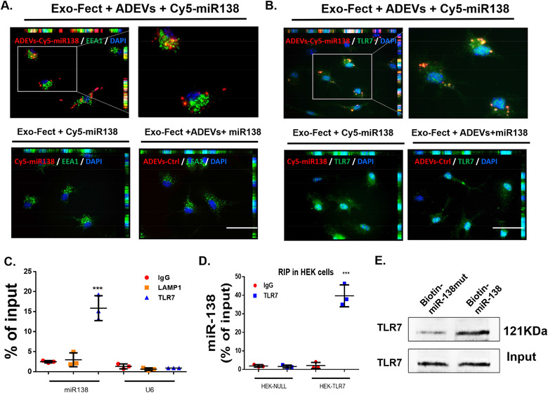
FIGURE 3.

ADEV miR‐138 interacts with murine TLR7 in the endosomes. (A) Representative fluorescence images of mouse primary microglial cells incubated with Exo‐Fect+ADEVs+Cy5‐miR138 or Exo‐Fect+Cy5‐miR138 (without ADEVs) or Exo‐Fect+ADEVs+miR138 (unstained) for 30 min followed by immunostaining of (A) an early endosome marker (EEA1, Green) and (B) TLR7 (Green). Exo‐fect+ADEVs+Cy5‐miR138, Cy5‐miR138 were loaded into ADEVs using by Exo‐Fect transfection kit; Exo‐fect+Cy5‐miR138, Cy5‐miR138 were loaded using by Exo‐Fect transfection kit (without ADEVs); Exo‐fect+ADEVs+miR138, unstained miR138 were loaded into ADEVs using Exo‐Fect transfection kit; Bars, 50 μm (n = 3). (C) TLR7 was immunoprecipitated from BV2 cells by IgG / TLR7 / LAMP1 antibody, followed by assessment of miR138 / U6 expression by real‐time PCR. One‐way ANOVA followed by Bonferroni's post hoc test was used to determine the statistical significance among multiple groups (n = 3). (D) TLR7 was immunoprecipitated from HEK‐Null / HEK‐TLR7 cells by IgG/TLR7 antibody, followed by assessment of miR138 expression by real‐time PCR. One‐way ANOVA followed by Bonferroni's post hoc test was used to determine the statistical significance among multiple groups (n = 3). (E) The protein of TLR7 were pull down by miR‐138‐biotin / miR‐mut‐138‐biotin with Streptavidin agarose beads in BV2 cells. All data are presented as mean ± SD or SEM of three individual experiments. *,P < 0.05; **,P < 0.01; ***,P < 0.001 versus control group.
