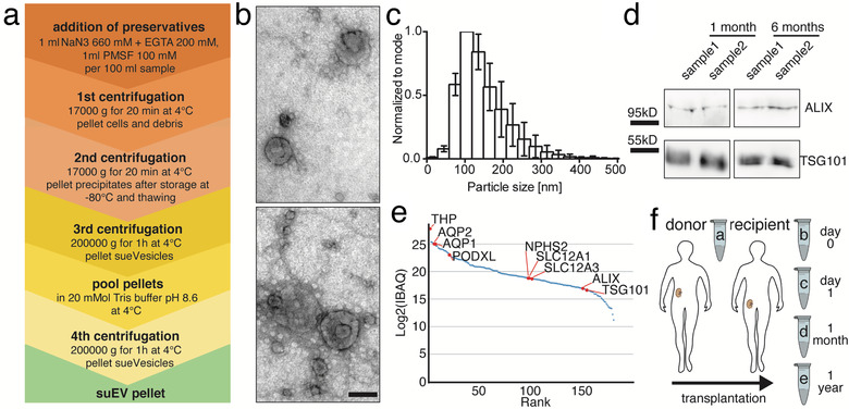
FIGURE 1.

Differential centrifugation separates small urinary extracellular vesicles (suEVs) originating from all segments of the nephron. Schematic overview of the employed protocol of differential centrifugation (a). Representative scanning electron microscopy of suEV pellets depicts EVs of typical exosomal cup shape and size and as well as smaller and bigger particles. Scalebar: 100nm (b). Size distribution of suEVs measured in 4 independent suEV samples of healthy volunteers measured by Nanoparticle Tracking, bin: 30nm, errorbars: SD, (c). Representative western blot analysis for exosomal markers ALIX and TSG101 in two separate suEV pellets after 1 and 6 months of storage in 8M Urea Buffer at −80°C (d). Mass spectrometry analysis suEV pellet is able todetectmarker proteins of all nephron segments. IBAQ: Intensity based absolute quantification (a.u.) (e) Schematic overview of the employed sampling protocol throughout living donor transplantation, a: donor sample; b: recipient sample day 0; c: recipient sample day 1; d: recipient sample 1 month; e: recipient sample 1 year (f)
