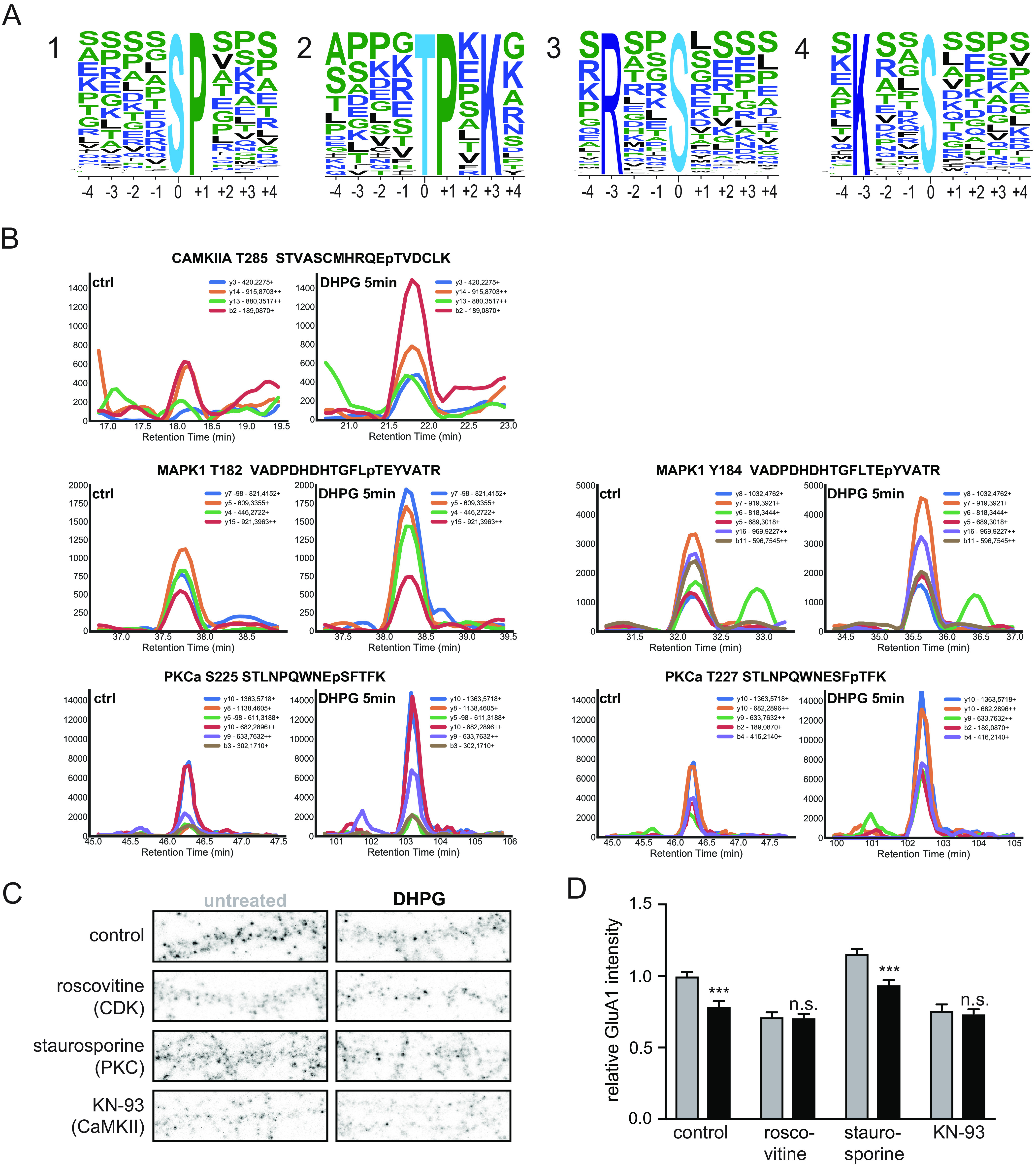
Fig. 4.

Kinases involved in DHPG-activated mGluR-LTD. A, MotifX sequence motifs of regulated phosphosites (p < 0.01) implicated in mGluR-LTD. B, Representative SRM traces of known kinases downstream of mGluR5 activation by DHPG. A clear increase in phosphorylation was observed after 5 min of DHPG stimulation compared with control for several activation sites of PKCa, CaMKIIa and MAPK1. C, Immunostaining of GluA1 subunits at the cell surface after control or DHPG treatment, pre-incubateed with control (untreated, n = 19)) or 3 different kinase inhibitors (Roscovitine (CDKs, n = 15), staurosporine (PKC, n = 12) and KN-93 (CaMKII, n = 12)). D, Relative quantification of cell surface GluA1 intensity; pre-incubation with roscovitine (n = 13) or KN-93 (n = 12) blocked DHPG-induced reduction in surface GluA1 levels in contrary to staurosporine (n = 12) pre-incubation which did not prevent mGluR-induced GluA1 internalization. Data are represented as mean±S.E. *** p < 0.001, n.s. not significant, as determined with a one-way ANOVA.
