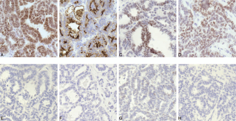
Figure 2.

Immunohistochemical staining of ovarian MLA. (A) PAX8 expression in the present case is strong and diffuse, which indicating the tumor arises from the female genital tract. (B) CD10 reveals cytoplasmic and luminal staining. (C) GATA3 positivity is helpful in confirming the diagnosis of mesonephric carcinoma, but the staining may be weak and/or focal. In this case, the staining of GATA3 is a little bit of weak and diffuse. (D) Immunohistochemical staining of TTF-1 is strong and diffuse. Immunohistochemistry showing totally negative staining with ER (E), PR (F), P53 (G), and P16 (H).
