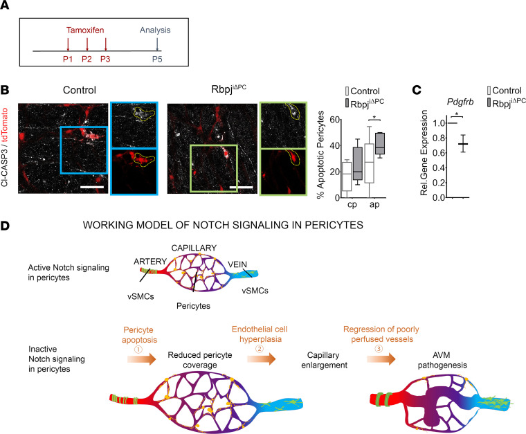
Figure 6. Increased pericyte apoptosis following perivascular inhibition of Notch signaling.
Control and RbpjiΔPC mice bred with Ai9 (TdTomato) reporter mice. (A) Offspring were treated with tamoxifen perinatally and analyzed for apoptosis at P5. (B) tdTomato reporter (red) utilized to represent pericytes in control and RbpjiΔPC vasculature. Anti–cleaved Caspase 3 (Cl-CASP3, white) labels apoptotic cells. Boxed regions show higher magnification of apoptotic pericytes. Quantification of pericyte apoptosis in capillary plexus (cp) and apoptotic pericytes (ap) displayed on the right (n = 5). (C) Gene expression of Pdgfrb in FAC-sorted retinal perivascular cells relative to β-actin. Box-and-whisker plots show median, minimum, and maximum values. Data were analyzed using unpaired 2-tailed t test with Welch’s correction. *P < 0.05. (D) Working model of AVM formation in RbpjiΔPC mice.

