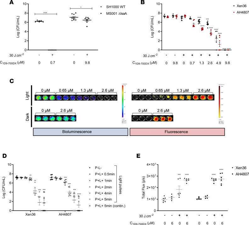
Figure 2. Photo-activated killing of S. aureus by 1D9-700DX.
(A and B) Photo-activated killing of S. aureus SH1000 WT versus MS001 ΔisaA (A) or Xen36 versus CA-MRSA AH4807 (B) grown to exponential phase (~1 × 10⁷ CFU/mL) upon treatment with 1D9-700DX or without photosensitizer (A), or step-wise increasing concentrations of 1D9-700DX (0.7–9.8 μM) (B). Bacteria were irradiated with red light at a radiant exposure of 30 J.cm–2 (+) or kept in the dark (–). (C) Bacterial bioluminescence (open emission filter, 10-second exposure) and fluorescence of 1D9-700DX (emission filter, Cy5.5; excitation, 640 nm; 10-second exposure) recorded with the IVIS Lumina II upon aPDT of S. aureus Xen36 or AH4807 (~1 × 10⁸ CFU/mL) with increasing concentrations of 1D9-700DX (0–2.6 μM). (D) Red light dose-response analysis of the killing of S. aureus Xen36 or CA-MRSA AH4807 grown to exponential phase and subjected to aPDT with 4.9 μM of 1D9-700DX or without photosensitizer. Bacteria were irradiated with red light for different periods of time (0.5–5 minutes) or kept in the dark (–). As a control, the bacteria were subjected to continuous (contin.) red light irradiation for 5 minutes. (E) H2O2 production upon aPDT of S. aureus Xen36 and AH4807 with 6 μM of 1D9-700DX or without photosensitizer. H2O2 was detected with 10 μM of an AquaSpark Peroxide Probe. In all experiments, irradiation was performed with a LED system that emits red light. Data are presented as mean ± SEM of 3 experiments performed in triplicates. Two-way ANOVA with subsequent Šidák multiple-comparison tests were used for statistical analysis. Significant differences compared with the negative control group (no 1D9-700DX and no light) are marked as follows: *P < 0.03; **P < 0.002; ***P < 0.0002; ****P < 0.0001.

