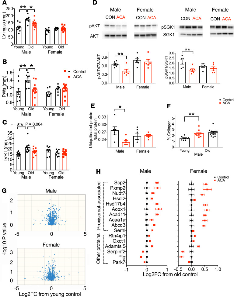
Figure 3. Effect of ACA on heart structure, function, signaling, and proteomic composition.
(A–C) Echocardiography was conducted on mice at 22 months of age (n = 8 per sex for old controls; n = 12 per sex for ACA treated), and included a set of young (6-month) controls (n = 8 per sex). (D and E) Analysis of cardiac signaling pathways and ubiquitinated protein was conducted on tissue samples from 25-month-old mice by Western blot. (F) The relative presence of collagen was assessed by Picrosirius red staining. (G) Proteomics analysis was conducted on samples from 25-month-old mice and 6-month-old young controls (n = 8 per sex for each treatment group). Volcano plots for each sex are shown, highlighting the lack of proteins that have a consistent change with age in protein abundance. (H) The log2 fold change in protein abundance from old control animals is shown for old mice treated with ACA for each protein that differs between control and ACA-treated mice after correction for FDR. Each error bar is a value for a different protein, with the figure showing relative change in abundance of proteins differing significantly between young and old mice on a control diet. Proteins were split according to their association with peroxisome, as defined by Gene Ontology (GO) cellular component annotation terms.*P < 0.05, **P < 0.01. In A–C and F, the P values were computed by running an ANOVA then Dunnett’s multiple comparison test, comparing young males and old ACA-treated males with old control males in separate comparisons. In D, P values were calculated with a Student’s t test.

