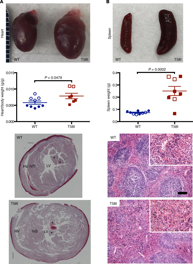
Figure 2. Organ enlargement in 6- to 8-month-old KrasT58I/+ mice.
(A) Gross appearance (top) and heart weight–to–body weight ratios (middle) of WT mice (n = 9, 7 male and 2 female mice) and their KrasT58I/+ littermates (n = 8, 6 male and 2 female mice). Mean ± SEM. Hearts were stained with Masson’s trichrome, and transverse sections were made at the papillary muscle level (bottom). RV, right ventricle; LV, left ventricle; IVS, interventricular septum. Asterisks indicate the papillary muscle. (B) Gross appearance (top), weights (mean and SEM are shown; middle), and representative histologic appearance (bottom) of representative spleens from WT and KrasT58I/+ mice. The mice analyzed were on either a F1 B6/129 (white circles or squares) or mixed B6/129 strain (colored circles or squares) background. Note expanded red pulp, with increased myeloid elements, in KrasT58I/+ spleens. Scale bar: 120 μM; 60 μM (for insets). Statistical significance was evaluated by Student’s t test.

