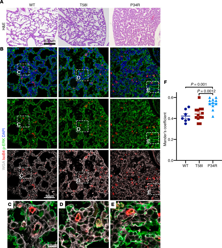
Figure 4. Lung morphology and phosphorylated ERK staining in neonatal mice.
(A) Representative neonatal lung tissue sections stained with H&E, showing poor lung inflation in CMV-Cre KrasLSL-P34R/+ (right) versus WT (left) or KrasT58I/+ (middle) neonates. (B) Immunohistochemical staining of representative neonatal lung sections with wheat germ agglutinin (WGA) (white), DAPI (blue), isolectin B4 (red), and a phosphorylated ERK (p-ERK) antibody (green) demonstrated a thickened septum in CMV-Cre KrasLSL-P34R/+ lungs. (C–E) p-ERK colocalizes with WGA in the neonatal lungs for all 3 genotypes, and CMV-Cre KrasLSL-P34R/+ lungs showed an increased degree of p-ERK colocalization with WGA (E), which was quantified in F. Scale bar: 10 μM (C–E); 50 μM (B); 400 μM (A).Statistical significance was evaluated by 1-way ANOVA with Tukey’s multiple comparisons test.

