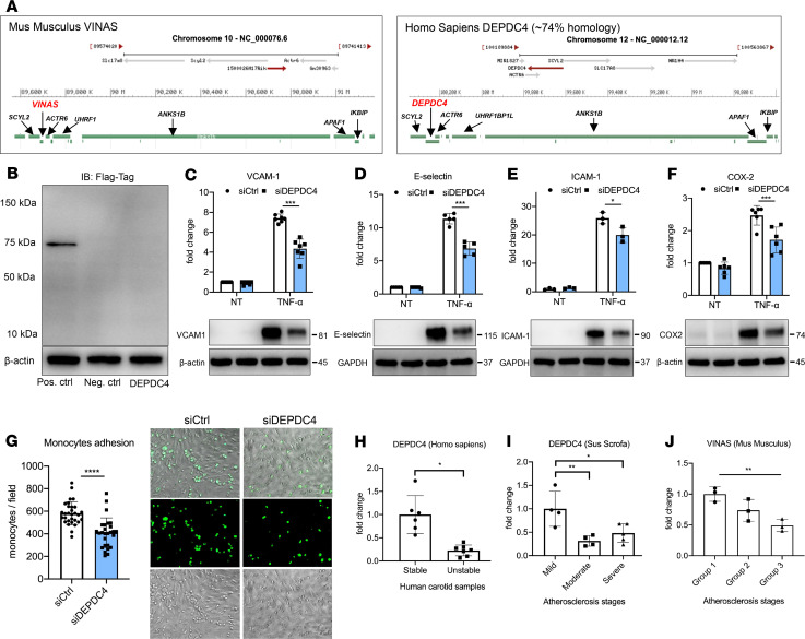
Figure 6. DEPDC4 is a human ortholog of VINAS.
(A) Illustration of the genomic locations of VINAS and DEPDC4 in the mouse and human chromosomes 10 and 12, respectively. (B) DEPDC4 does not encode for a protein or peptide. To test the coding potential, DEPDC4 sequence was cloned upstream of the 3xFlag-Tag cassette, transfected in HEK293T cells, and immunoblotted for Flag antibody; positive control was provided with the kit (n = 3 experiments). DEPDC4 silencing decreases the protein expression of VCAM-1 (C, n = 7), E-selectin (D, n = 5), and ICAM-1 (E, n = 3) COX-2 (F, n = 6) in HUVECs activated with 20 ng/mL TNF-α. (G) DEPDC4 knockdown decreases THP-1 monocyte adhesion to HUVEC monolayers activated with TNF-α for 4 hours (5 ng/mL, representative images and quantification of adhered monocytes). (H) RT-qPCR of DEPDC4 in human carotid arteries with stable (n = 6) or unstable (n = 7) atherosclerotic plaques. Scale bar: 50 μm. (I) Expression of DEPDC4 from RNA-Seq analyses of lesions with increasing severity of coronary atherosclerosis in Yorkshire pigs fed an HCD for 60 weeks (n = 4/group). (J) RT-qPCR of VINAS expression in aortic intima of LDLR–/– mice at 0, 2, and 12 weeks of an HCD (n = 3/group). Data represent the mean ± SD. Statistical differences were calculated using unpaired 2-tailed Student’s t test except for multiple comparisons (I and J) in which 1-way ANOVA with Bonferroni’s correction was used. *P < 0.05, **P < 0.01, ***P < 0.001, ****P < 0.0001.

