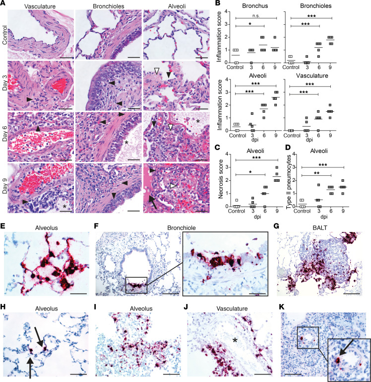
Figure 2. Histopathological changes and detection of SVV ORF63 RNA in lung tissue of SVV-infected cynomolgus macaques.
(A–D) H&E-stained lung sections (A) were scored for inflammation (B), necrosis (C), and abundance of type II pneumocytes (D). *P < 0.05, **P < 0.01, ***P < 0.001 by 1-way ANOVA and Bonferroni’s correction. (A) Black arrowheads, inflammatory cells; white arrowheads, necrosis; asterisks, fibrin deposition; arrow, edema. Scale bar: 50 μm. (E–K) Lung sections stained for SVV ORF63 RNA by ISH (purple) and counterstained with hematoxylin (blue) at 3 dpi (E–G), 6 dpi (H–J), and 9 dpi (K). Enlargement of areas indicated by black boxes are shown. Scale bars: 50 μm (E and F, left panel), 100 μm (H–K), 200 μm (F [right panel] and G). Arrows, SVV RNA+ cells; asterisk, blood vessel; BALT, bronchus-associated lymphoid tissue.

