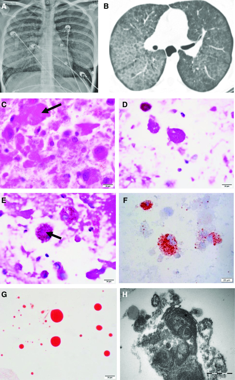
Figure 1.
(A) Frontal chest X-ray with bilateral perihilar hazy air space opacification. (B) High-resolution chest computed tomographic scan showing interlobular septal thickening forming a crazy paving pattern. (C) Hematoxylin and eosin showing extracellular eosinophilic hyaline material (arrow). (D) Periodic acid–Schiff with diastase–positive extracellular globular material. (E) Periodic acid–Schiff with diastase–positive granular deposit in intracytoplasmic macrophages (arrow). (F) Oil red O stain highlighting intracytoplasmic (macrophages) lipid vacuoles. (G) Oil red O stain–positive extracellular hyaline globules. (C–G) Scale bars, 20 μm. (H) Electron microscopy showing myelin-like whorled membranous structures (lamellar bodies) (scale bar, 0.5 μm).

