. 2020 Nov 16;9:e59157. doi: 10.7554/eLife.59157

Figure 1. CRMs controlling Ciinte.Msx expression in VDML.

(A) Snapshot of the Ciinte.Msx locus depicting ATAC-seq profile at mid-neurula stages, tested genomic regions, transcript models and conservation between C. robusta and C. savignyi (from https://www.aniseed.cnrs.fr/ and Dardaillon et al., 2020; Madgwick et al., 2019). (B–E) Representative examples of X-gal stained embryos at late gastrula stages (B, D) and early tailbud stages (C, E) following C. intestinalis embryos electroporation of Ciinte.Msx-Intergenic (B, C) and Ciinte.Msx-up1 (D, E). Embryos are shown in dorsal view (B, D) and in lateral view with dorsal to the top (C, E), and anterior to the left. Scale bar: 100 μm. (F) Schematic representation of the various constructs and their activity at early tailbud stages in DML (blue) and VML (purple) (n indicates the total number of embryos examined; N indicates the number of independent experiments).

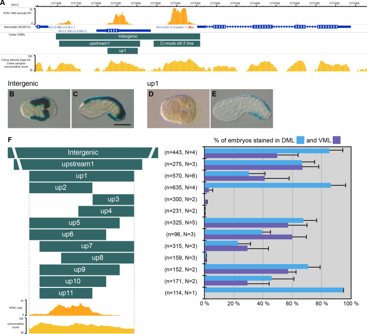
Figure 1.

Figure 1—figure supplement 1. Developmental regulators of caudal PNS in C. intestinalis.

Figure 1—figure supplement 1.

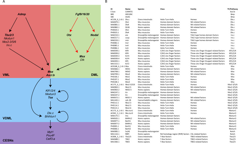
(A) Provisional GRN for caudal PNS specification in C. intestinalis. The genes depicted and their interactions are based on previous publications (Roure et al., 2014; Pasini et al., 2006; Roure and Darras, 2016; Waki et al., 2015; Joyce Tang et al., 2013; Bertrand et al., 2003) and hypotheses described in the Materials and methods section. Genes whose requirement for caudal PNS formation has been shown by loss-of-function are in bold. Demonstrated direct regulations are shown in red. (B) List of matrices and consensus motifs used to identify TFBS in CRMs.
Figure 1—figure supplement 2. Model for Ciinte.Msx regulation.

Figure 1—figure supplement 2.

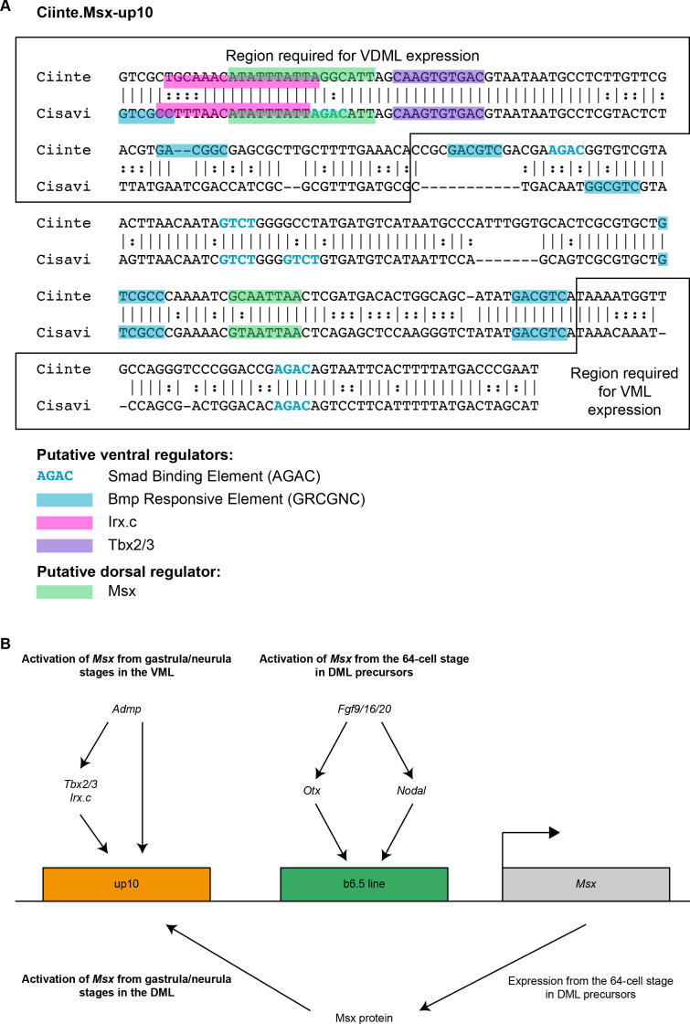
(A) Identification of putative TFBS for candidate upstream factors in Ciinte. Msx-up10 aligned with its counterpart from C. savignyi. All putative sites for ventral factors (SBE, BRE, Tbx2/3, Nkxtun1, Nkx2-3/5/6 and Irx.c) and dorsal factors (Msx and Su(H)/Rbpj) have been mapped, but only conserved sites are shown: 4 BRE, 3 SBE, 1 Irx.c, 1 Tbx2/3 and 2 Msx. The region deleted in up2 and up11 abuts a BRE and contains a SBE; the loss of these sites might thus be responsible of the absence of activity in the VML. The region deleted in up8 and required for VDML activity contains 1 BRE, 1 Irx.c, 1 Tbx2/3 and 1 Msx sites. Note that the size of the highlighted site for of a given TF may vary depending on the matrix used. (B) Working model for Msx transcriptional regulation in the VDML. Msx expression is initiated in DML precursors at the 64-cell stage (Roure et al., 2014) through the proximal/early CRM (denoted b6.5 line, green) activated by Otx and Nodal (via Smad2/3). The Msx protein produced via this regulation would activate Msx transcritption in the DML at gastrula/neurula stages via the distal/late CRM (denoted up10, orange). The same distal CRM would activate Msx expression in the VML directly by Bmp signaling activated by the Admp ligand (via the SBEs and BREs) or Admp targets (Irx.c and Tbx2/3).