. 2020 Nov 16;9:e59157. doi: 10.7554/eLife.59157

Figure 8. Overall conservation of midline gene expression in Stolidobranchia ascidians.

(A) Phylogenetic relationships between C. intestinalis, P. mammillata, M. appendiculata and H. roretzi (estimated divergence times come from Delsuc et al., 2018). (B–M) In situ hybridization for Msx (B, H) at neurula stages, for Ascl.b (C, I), Klf1/2/4 (D, J), Nkxtun3 (E, K), Tox (F, L), and Dlx.c (G, M) at tailbud stages in embryos of M. appendiculata (B–G) and H. roretzi (H–M). Note that all genes except Moappe.Dlx.c are expressed in tail midlines. Embryos are shown in lateral view with dorsal to the top and anterior to the left. Scale bars: 100 μm.

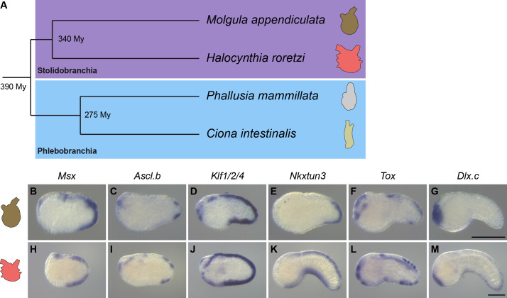
Figure 8.

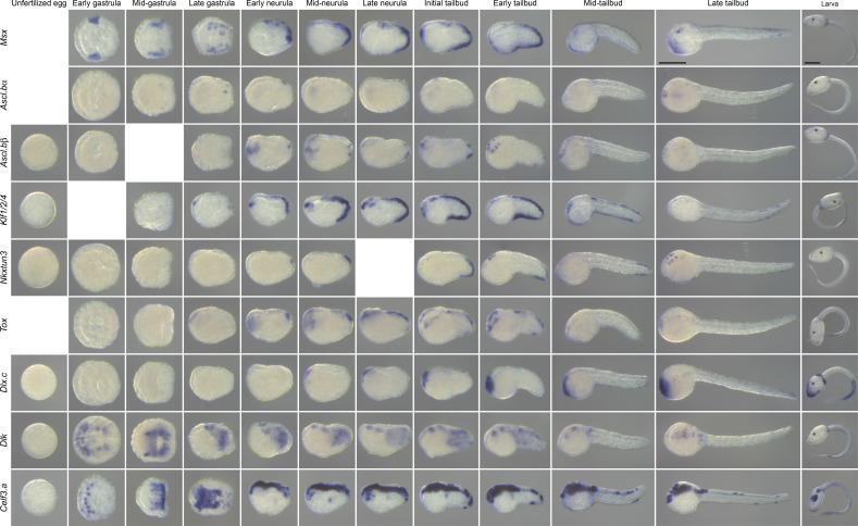
Figure 8—figure supplement 1. Expression pattern of caudal PNS genes in M. appendiculata.

Figure 8—figure supplement 1.

In situ hybridization results at several developmental stages for the M. appendiculata orthologs of the C. intestinalis caudal PNS genes: Msx, Ascl.b (Ascl.bα and Ascl.bβ), Klf1/2/4, Nkxtun3, Tox, Dlx.c, Dlk, and Celf3.a. Scale bars: 100 μm.
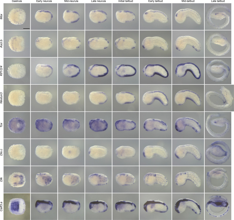
Figure 8—figure supplement 2. Expression pattern of caudal PNS genes in H. roretzi.

Figure 8—figure supplement 2.

In situ hybridization results at several developmental stages for the H. roretzi orthologs of the C. intestinalis caudal PNS genes: Msx, Ascl.b, Klf1/2/4, Nkxtun3, Tox, Dlx.c, Dlk, and Celf3.a. Scale bar: 100 μm.
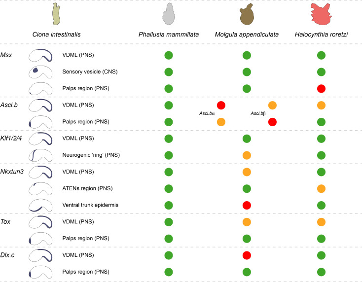
Figure 8—figure supplement 3. Comparison of midline TFs expression in C. intestinalis, P. mammillata, M. appendiculata, and H. roretzi.

Figure 8—figure supplement 3.

For each gene, the main sites of expression from late neurula stages in C. intestinalis (Roure and Darras, 2016; Imai et al., 2004) are schematized on the left. For the other species, identical pattern is represented as a green circle, expression in part of the tissue as an orange circle, and absence of expression as a red circle. For simplicity, developmental timing has not been described, but it has been taken into account for comparisons. For example, Ciinte.Ascl.b is expressed in the palps forming region from initial tailbud to late tailbud stages, while Moappe.Ascl.bα was detected in the palps only at late tailbud stages. This difference was depicted by an orange circle.