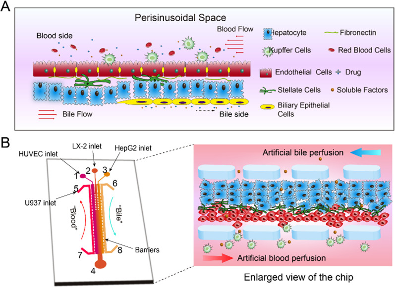
FIG. 1.
Schematic diagram of the liver-on-a-chip design. (a) Microphysiological structure of hepatic sinusoids in vivo including cell composition, spatial arrangement, blood flow, and bile duct formation; (b) Schematic diagram of the liver-on-a-chip: (1) HUVEC cells inlet, (2) LX-2 cells inlet, (3) HepG2 cells inlet, (4) outlet, (5) and (7) artificial blood channel, and (6) and (8) artificial bile channel (enlarged for real cell arrangement in the chip). The central channel was 1 cm in length, 200 μm in width, and 100 μm in height. The side perfusion channels were 1 cm in length, 100 μm in width, and 100 μm in height. The gap between the micro-fences was 20 μm.

