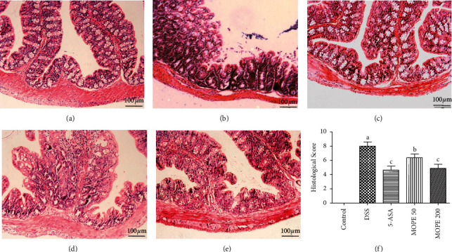
Figure 3.

Effects of MOPE on the histopathological characterization of DSS-colitis mice. Representative HE-stained sections of the distal colonic tissues from the (a) control, (b) DSS, (c) 5-ASA (50 mg/kg), (d) MOPE (50 mg/kg), and (e) MOPE (200 mg/kg) groups. All images were acquired using 200× magnification. (f) Histological scores of colonic abnormalities. Values with different letters (a-c) differ significantly (p < 0.05).
