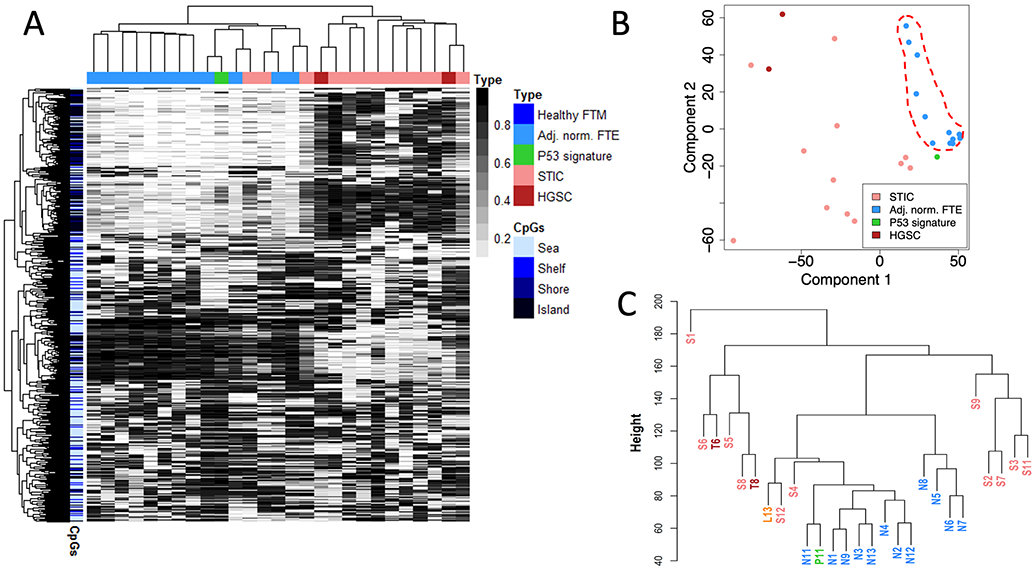
Figure 2. Unsupervised analysis of genome-wide methylation of HGSC-precursor lesions of the fallopian tube.

(A) Methylation heatmap showing the 1000 most variable CpG probes as determined by MethylationEPIC analysis. (B) Corresponding multidimensional analysis showing relative clustering of adjacent-normal epithelia (dashed red line). (C) Sample dendrogram showing that the majority of adjacent-normal epithelia and p53 signature lesion are readily distinguishable from STIC lesions and HGSC tumors. STIC-HGSC pairs are shown in bold red. FTM – Fallopian tube mucosa, FTE – Fallopian Tube Epithelium
