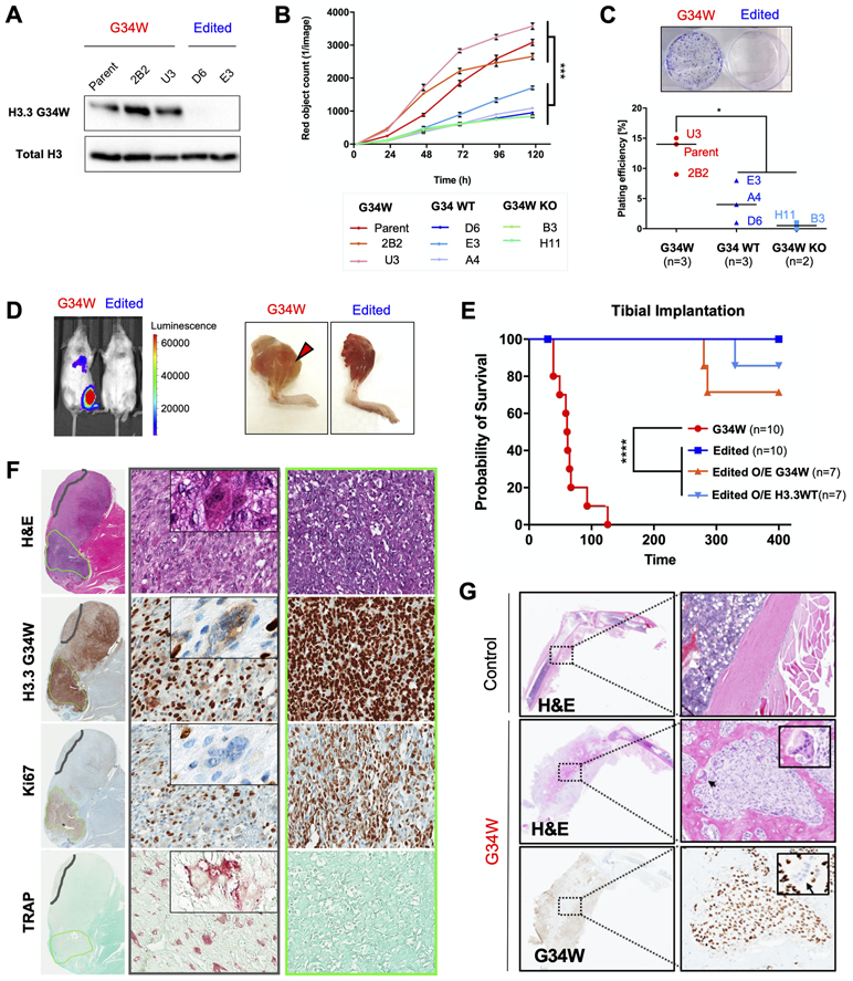
Figure 1. G34W is necessary for tumourigenesis and promotes aggressive osteolytic bone lesions in an orthotopic xenograft model.

(A) G34W immunoblotting of Im-GCT-4072 G34W (Parent: n=1; Clone: n=2) and edited (Repair to WT: n=2) lines.
(B) G34W lines (Parent: n=1; Clone: n=2) of Im-GCT-4072 proliferate faster than edited lines (Repair to WT: n=3; G34W-KO: n=2), as measured using the IncuCyte live-cell analysis system for 5 consecutive days. Data are presented as mean red object count ± SD from five technical replicates per line. Statistical significance assessed using Student’s t-test based on averaged observations from biological replicates (independent CRISPR clones, labeled).
(C) Representative images of G34W (left) and edited (right) cell colonies in culture plates (top panel). G34W lines (Parent: n=1; Clone: n=2) of Im-GCT-4072 exhibit increased colony formation relative to edited lines (Repair to WT: n=3; G34W KO: n=2), as measured by manual counting of colonies stained with crystal violet after 3 weeks (bottom panel). Two technical replicates were counted per line, and data are presented as an average of biological replicates (independent CRISPR clones, labeled). Statistical significance assessed using Student’s t-test.
(D) Left: Representative bioluminescence Xenogen IVIS 200 imaging of mice implanted in the tibia with Im-GCT-4072 G34W and edited luciferase-tagged lines.
Right: Representative images of mice tibias implanted with G34W and edited cells after skin removal at the time of sacrifice.
(E) Kaplan-Meier survival curve for orthotopic tibial implantation of Im-GCT-4072 G34W (Parent: n=1, Clone: n=2; n=10 mice), edited lines (Repair to WT: n=2, n=10 mice), edited O/E G34W (n=7 mice), edited O/E H3.3WT (n=7 mice) in NRG mice illustrates the dependence of tumour formation on the presence of G34W mutation.
(F) H&E, and H3.3G34W, TRAP (tartrate-resistant acid phosphatase) and Ki67 IHC for a representative tibial xenograft tumour derived from implantation of Im-GCT-4072 G34W parental cells. 20X (70μm) magnified area illustrates a histological compartment (middle panel) with differentiated stromal cells and abundant TRAP+ osteoclasts (an example of giant multinucleated osteoclast is featured in the inset). Right panel illustrates a histological compartment with undifferentiated stromal cells, high Ki67 staining, and absence of TRAP+ osteoclasts.
(G) Representative H&E and G34W IHC of decalcified legs derived from tibial implantation of Im-GCT-6176 G34W parental cells, illustrating the osteolytic effect of G34W stromal cells relative to a control contralateral leg. Inset features reactive G34W-negative osteoclasts observed at the interface between G34W-positive neoplastic stromal cells and normal bone.
