
Figure 2. Investigation of patient 11-272 carrying the SHOC1 variant.
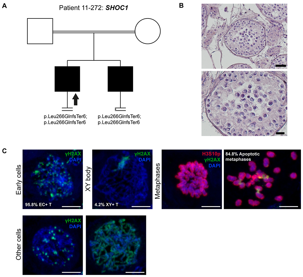
A) The pedigree structure shows the segregation of the SHOC1 variant (p.Leu266GlnfsTer6). B) H&E staining of histological sections from the testis biopsy of the patient carrying the SHOC1 variant. Scale bar on the upper image represents 50 μm and on the lower image 20 μm. C) Immunofluorescent staining of histological sections from the testis biopsy of the patient’s brother carrying the SHOC1 variant using γH2AX (Green), H3S10p (red), and DAPI (blue). Scale bar represents 5μm. Early cells were observed in 95.8% of the round tubules that were counted. Extremely reduced XY body positive tubules were observed, indicating that the cells rarely reach the pachytene stage. A high density of metaphases was observed (29 metaphases/mm2) with a high percentage of apoptotic metaphases (84.8%). The organization of the apoptotic metaphases appeared to be more chaotic, as the chromosomes within these metaphases were dispersed. The early cells displayed an aberrant pattern of γH2AX spots. Some cells displayed an aberrant pattern whereby γH2AX is localized on the entire axis of the DNA strands.