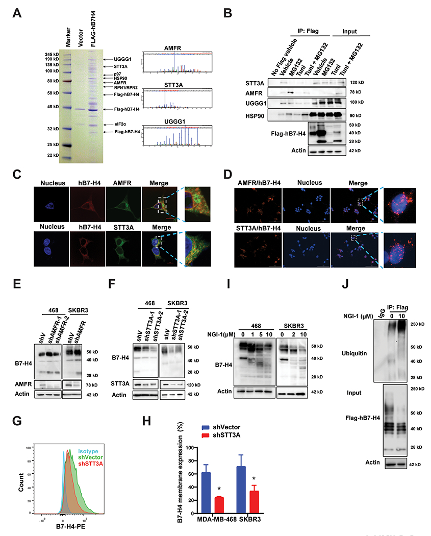
Figure 3. Identification of the E3 ligase and glycosyltransferases of B7-H4 that govern B7-H4 protein stability and function.

(A) Stable expression of Flag-hB7-H4 was engineered to MDA-MB-468 cells (MDA-MB-468-Flag-hB7-H4). B7-H4 complex was then purified followed by mass spectrometry analysis. Coomassie blue staining of the purified B7-H4 immunocomplex is shown. The ubiquitin E3 ligase AMFR and several glycosyltransferases including STT3A, RPN1, RPN2, and UGGG1 were identified, and the representative spectra of AMFR, STT3A and UGGG1 are shown. (B) Validation of biochemical interactions of B7-H4 with AMFR, STT3A, UGGG1 as well as HSP90. MDA-MB-468-Flag-hB7-H4 cells were utilized for immunoprecipitation using anti-Flag M2-beads in the presence or absence of MG132 and/or tunicamycin. The interactions of AMFR, STT3A, UGGG1 as well as HSP90 with B7-H4 were measured by immunoblot. (C) Double immunofluorescence staining hB7-H4-Flag with AMFR or STT3A in MDA-MB-468-Flag-hB7-H4 cells followed by the confocal microscope (Scale bar =10 μm). (D) MDA-MB-468-Flag-hB7-H4 cells were subjected to duolink in situ PLA assay with specific Flag mouse antibody and AMFR or STT3A rabbit antibody (Scale bar = 100 μm). Red dots indicate the binding of the indicated two proteins. (E) AMFR knockdown results in upregulation of B7-H4. Stable knockdown of AMFR in MDA-MB-468 and SKBR3 were established. The expression of AMFR and B7-H4 were examined by immunoblotting. (F) STT3A knockdown leads to downregulation of B7-H4. STT3A stable knockdowns in MDA-MB-468 cells were established. The expression of STT3A and B7-H4 were examined by immunoblotting. (G) Decreased membrane B7-H4 in STT3A knockdown cells. MDA-MB-468-shVector, MDA-MB-468-shSTT3A, SKBR3-shVector, and SKBR3-shSTT3A cells were stained with PE anti-human B7-H4 antibody followed by flow cytometry. Representative images are shown. (H) The quantification of membrane staining of B7-H4 in MDA-MB-468-shVector, MDA-MB-468-shSTT3A, SKBR3-shVector, and SKBR3-shSTT3A cells are shown. (I) MDA-MB-468 and SKBR3 cells were treated with 10 μM OST inhibitor NGI-1 for 24 h. The expression of B7-H4 was examined by immunoblotting. (J) Blockade of B7-H4 glycosylation by NGI-1 enhances B7-H4 ubiquitination. 293T cells were transfected with Flag-hB7-H4 in the presence or absence of 10 μM NGI-1 for 24 h. Then Flag-hB7-H4 was immunoprecipitated followed by immunoblotting using antibody against ubiquitin.
