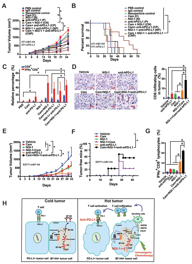
Figure 7. The anti-tumor efficacy of the combination of NGI-1 and a doxorubicin analog are enhanced by PD-L1 blockade.

(A) 4T1-hPD-L, where the basal mouse PD-L1 was knocked out followed by adding back human PD-L1 gene, and 4T1-hB7-H4 cells were orthotopically injected into the left fourth mammary fat pad and allow to grow around 100 mm3, followed by injection of Cam (25 mg/kg, i.p.) for 4 times, NGI-1 nanoparticle (10 mg/kg, i.v.) as well as PD-L1 antibody durvalumab (5 mg/kg, i.p.) for 3 times. The tumor growth was monitored twice per week. n=8 mice per group. (B) Survival curve of the mice of the combination of Cam, NGI-1 nanoparticle and durvalumab. n=8 mice per group. (C) On day 18, mouse tumors were harvested, and digested followed by flow cytometry of staining IFNγ and CD8. Quantification of IFNγ+CD8+ or CD8+ cell in the tumor mass is shown. n=3 mice per group. (D) Tumor tissues were subjected to the immunohistochemically staining with anti-CD8 antibody. n=3 mice per group. The representative images are shown. Scale bar, 20 μm. Quantification of CD8+ infiltrating cells is shown. (E) E0771-mB7-H4 cells (1×10⁵) were orthotopically injected into the left fourth mammary fat pad. When the tumors were visible on day 6, Cam (5 mg/kg, i.p.) was injected on day 6, 8, 10, and 12; NGI-1 nanoparticles (10 mg/kg, i.v.) were injected on day 7, 9, 11, and 12; and the anti-mPD-L1 antibody (5 mg/kg, i.p.) was injected on day 8, 10, and 12. The tumor growth was monitored twice per week. n=9 mice per group. (F) The percentage of tumor free mice was evaluated every week for 42 days (n=9). Log-rank (Mantel-Cox) test was performed for statistical analysis. (G) On day 27, spleens were harvested followed by flow cytometry of staining IFNγ and CD8. Quantification of IFNγ+CD8+ or CD8+ cells in the spleens is shown. n=3-5 mice per group. (H) The proposed working model of the combination of immunogenic chemotherapy, NGI-1 and PD-L1 blockade.
