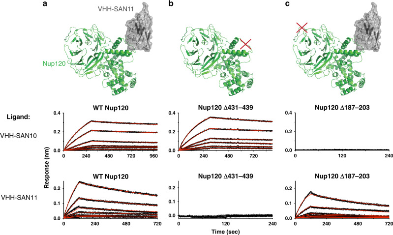
Fig. 4. Mutational analysis of Nup120 confirms the binding site of VHH-SAN10.
Bio-layer interferometry (BLI) showing association and dissociation kinetics for VHH-SAN10 and VHH-SAN11 with Nup120 mutants. Nanobodies with a biotinylated C-terminal Avi-tag were fixed (ligand) and nups used as analytes. Curves were corrected for buffer background. Each set of curves is a twofold dilution series from 10 nM analyte. Data are indicated by the black dotted lines and the red lines show the globally fitted curves. a Structure of Nup1201–757-VHH-SAN11 and BLI data of wild type (WT) Nup1201–757 as the analyte. b Illustration and BLI data of Nup1201–757 ∆431–439 (GSSx3) as the analyte. c Illustration and BLI data of Nup1201–757 ∆187–203 (GGSx5) as the analyte.

