Fig. 3.
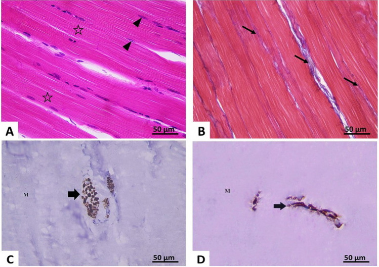
Photomicrographs of section of right gluteal muscle of the control group X400 A H&E-stained section showing well-defined cross striations (*) and peripheral elongated nuclei (▲).B Mallory’s trichrome: thin bundles of collagen fibers (↑) are seen in the interstitium between the muscle fibers. C, D Immune-histochemical reaction for neurofilament light chain: showing positive expression (↑) in transverse (C) and longitudinal nerves (D) between muscle fibers (M)

